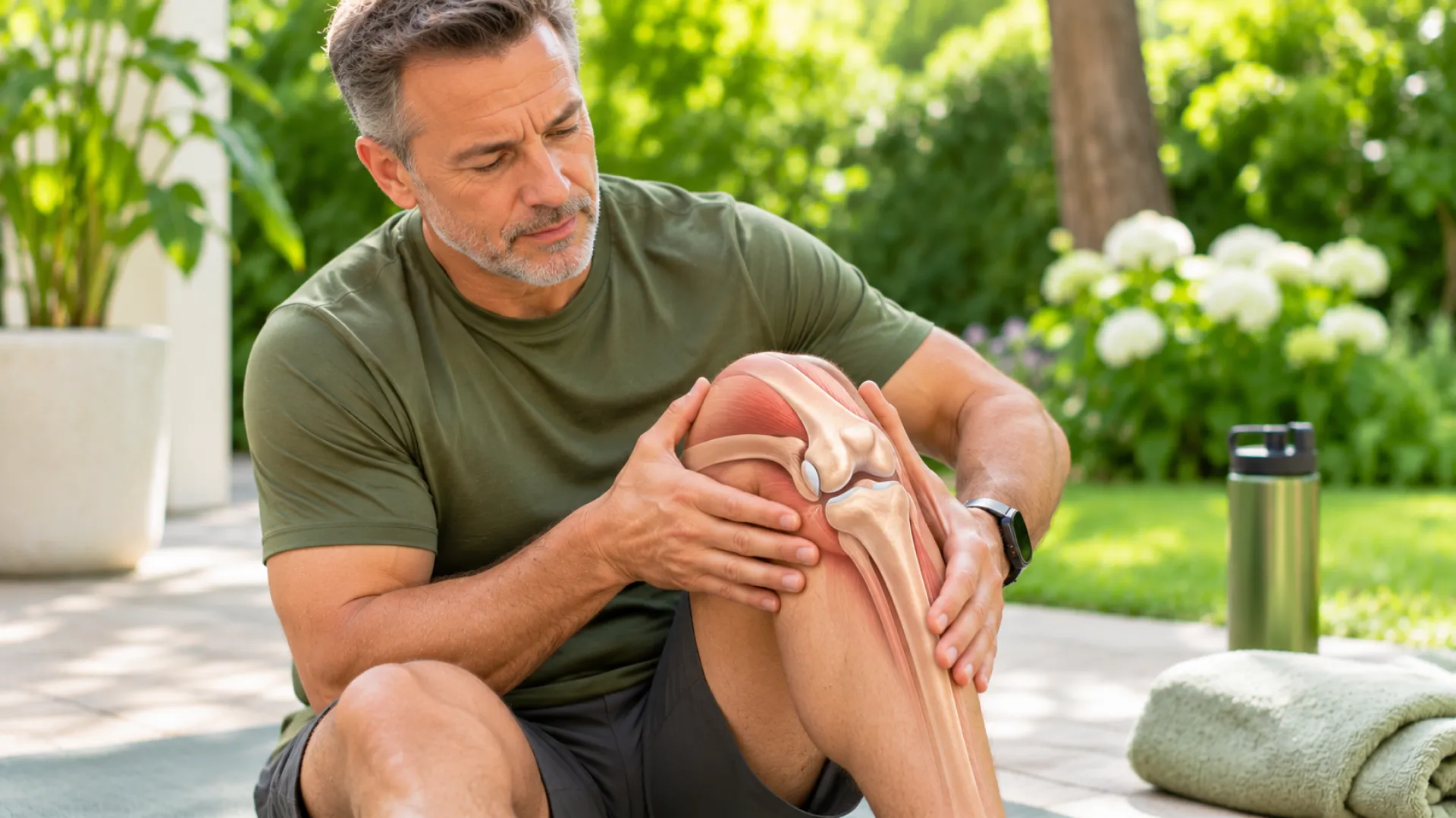
You don’t think about your knees much. Until one hurts.
Then the doctor starts saying words like ACL, meniscus, cartilage. Patella. And you nod along, not totally sure what any of it means.
That’s normal. Knee anatomy sounds hard, but it’s really just a few bones, some straps, some cushions, and the muscles that move it all.
So, let us now look into “What Is Knee Anatomy and How Does the Knee Joint Work?”!
Knee Anatomy Overview
The knee is the biggest joint in your body. Thigh on top, lower leg on the bottom, and a lot going on in between.
Most people call it a hinge. Close, but not quite. A door hinge only swings one way. Your knee bends and straightens, sure, but it also turns and slides a little as it moves. That’s how you can pivot, squat, and change direction without falling over.
It’s actually two joints working as one. The main one, where the thighbone sits on the shinbone, takes your weight. The other is where your kneecap slides in a groove on the front of the thighbone.
And it takes a lot of load. Researchers put sensors inside knee implants and found the knee carries more than 2 times your body weight in almost everything you do. Around 2.5 times for most daily tasks. Stairs were the worst.
So a 150-pound person might be putting close to 375 pounds through one knee on the stairs. No wonder small problems turn into real pain.
Bones of the Knee Anatomy
When you look at the knee in anatomy, it starts with bones. Three make the joint. One sits right next door.
Femur (Thighbone)
Longest, strongest bone you have. Runs from your hip to your knee.
At the bottom, it splits into two rounded knobs, one on the inside and one on the outside. Doctors call them condyles. These knobs roll and slide on top of your shinbone every time you bend. Between them, at the front, is a groove where the kneecap rides.
Tibia (Shinbone)
The big bone down the front of your lower leg. You can feel it right under the skin.
Its top is fairly flat. The thighbone’s two knobs rest on it, and most of your weight passes through here. Just below the knee, there’s a bump you can feel. The tendon from your kneecap hooks on right there.
Patella (Kneecap)
Small. Flat-ish. Shaped a bit like a triangle.
The weird thing about the kneecap is that it sits inside a tendon. It works like a lever, giving your thigh muscles extra pull when you straighten your leg. Without it, standing up from a chair would take a lot more effort.
The back of it is lined with some of the thickest cartilage in the whole body. It needs it. Squatting and stairs put huge force on the kneecap.
Fibula
Thin bone on the outer side of your lower leg.
Not part of the knee joint, technically. Carries hardly any weight. But it still matters, because a knee ligament and one of your hamstring tendons both anchor to the top of it.
Parts of the Knee: Ligaments
Ligaments hold bone to bone. Tough, short bands. Think strong tape that keeps the joint from sliding where it shouldn’t.
Four main ones in the knee. Two cross through the middle. Two run down the sides.
ACL (Anterior Cruciate Ligament)
The famous one.
It runs through the middle of the knee and stops your shinbone from sliding forward. It also keeps twisting under control. “Cruciate” just means cross-shaped. The ACL and PCL cross each other like an X.
About 200,000 ACL tears happen in the US every year. Most don’t come from a hit. A sudden stop. A fast cut on the court. Landing a jump the wrong way. People often say they heard a pop, then the knee swelled up fast.
PCL (Posterior Cruciate Ligament)
Sits right behind the ACL. Bigger. Stronger. Gets hurt a lot less.
It keeps your shinbone from sliding backward. The classic PCL injury is a car crash where the knee slams into the dashboard. Or a fall straight onto a bent knee.
MCL (Medial Collateral Ligament)
Runs down the inner side of the knee.
Something hits the outside of your knee, the knee buckles inward, and the MCL takes the hit. Football and soccer players know this one well. Good news: most MCL sprains heal without surgery.
Pain on the inside of the knee can have other causes too. We cover them in our post on inner knee pain.
LCL (Lateral Collateral Ligament)
The MCL’s partner on the outer side. Thinner and injured less often, usually from a blow to the inside of the knee that pushes it outward.
Parts of the Knee: Tendons
Tendons are different. They tie muscle to bone. Muscle pulls, tendon pulls the bone, joint moves.
Patellar Tendon
From the bottom of your kneecap down to that bump on your shin.
Strictly speaking, it links bone to bone, so some doctors call it a ligament. Whatever you call it, it does a tendon’s work, passing the pull of your thigh muscles down to straighten your leg.
Too much jumping or running and it gets sore right below the kneecap. That’s jumper’s knee.
Quadriceps Tendon
Sits above the kneecap. Connects your big front thigh muscles to it.
Picture a chain. Thigh muscles, quad tendon, kneecap, patellar tendon, shin. Pull on one end, and your leg straightens. That’s the whole system you use to stand up, kick, or climb.
Tears here are rare. When they happen, it’s usually someone over 40 after a fall.
Hamstring Tendon
Back of the knee. Bend your leg a little, and you can feel them as cords on both sides.
These connect the hamstring muscles to your shinbone and fibula, and they bend your knee. They also back up the ACL by holding the shin from sliding forward. Fun fact: surgeons often borrow a strip of hamstring tendon to rebuild a torn ACL.
Parts of the Knee: Cartilage and Menisci
Two kinds of cushion in the knee. They’re not the same thing, and people mix them up all the time.
Articular Cartilage
This is the smooth, shiny coating on the ends of your bones. Thighbone, shinbone, back of the kneecap. If you’ve ever seen the white glossy tip of a chicken bone, that’s basically it.
It lets the bones glide with almost no friction.
The problem? It has no blood supply. So once it’s damaged, it barely heals.
When it wears down slowly over the years, that’s osteoarthritis. When it’s worn all the way through and bone touches bone, doctors call it bone-on-bone knee. Morning stiffness is often the first hint, and it’s worth reading up on joint stiffness if that sounds familiar.
Medial and Lateral Meniscus
Two C-shaped pads of tough, rubbery cartilage sitting between the thighbone and shinbone. One on the inside (medial), one on the outside (lateral).
They soak up shock. They spread your weight out so it doesn’t land on one small spot. And they cup the end of the thighbone so the knee stays steady.
The inner one is stuck down more firmly, so it can’t move out of the way as well. That’s why it tears more often.
Only the outer rim of each meniscus gets blood. Tears out there can sometimes heal. Tears deeper in usually don’t.
Here’s something most people don’t know. Meniscus tears are super common, and a lot of them don’t hurt at all. A big MRI study of almost 1,000 adults over 50 found 31% had a tear. Past age 70, it was over half. And about 55% of those tears caused no symptoms whatsoever.
Plenty of people just live with one. On a forum thread about skipping meniscus surgery, one guy said he’d had his tear for 35 years. It locked up now and then. He never thought the downtime from surgery was worth it. Another person did two months of physical therapy, still got the odd twinge, and planned to only go for surgery if things got worse.
Parts of the Knee: Muscles
Muscles aren’t inside the joint. But they’re its bodyguards. Strong muscles take load off your ligaments and cartilage. Weak ones leave the joint to fend for itself.
Quadriceps
Four muscles on the front of your thigh. Rectus femoris down the middle, vastus lateralis on the outside, vastus medialis on the inside just above the knee, and vastus intermedius tucked underneath.
They straighten the knee and help keep the kneecap tracking straight in its groove.
Weak quads go hand in hand with more knee pain and faster cartilage wear. That’s why almost every knee rehab plan starts with them.
Hamstrings
Three muscles down the back of the thigh. Biceps femoris on the outside. Semitendinosus and semimembranosus on the inside.
They bend your knee and help the ACL do its job. If your hamstrings are a lot weaker than your quads, ACL injury risk goes up.
Your calf muscle crosses the back of the knee too. And your glutes, up at the hip, decide whether your knee caves inward when you land. Not knee muscles exactly. Still part of the story.
Bursae of the Knee Anatomy
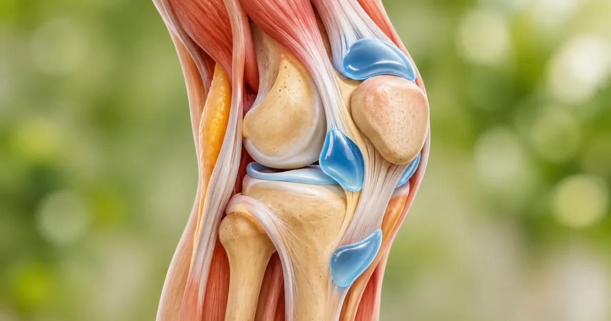
Bursae are tiny fluid sacs. Little water cushions sitting between bone and whatever slides over it. Your knee has more than ten of them.
Most never cause trouble. A few do:
- Prepatellar bursa, in front of the kneecap. Swells up in people who kneel a lot. Old name: “housemaid’s knee.”
- Infrapatellar bursa, just below the kneecap. Kneeling and jumping bother it.
- Pes anserine bursa, inner side, a little below the joint. Runners get it, and so do people with knee arthritis.
- Suprapatellar bursa, above the kneecap. It’s connected to the joint, so it puffs up when the knee makes extra fluid.
A sore, swollen bursa is called bursitis.
Then there’s the Baker’s cyst. A pocket of fluid behind the knee, usually from extra joint fluid pushed back there by arthritis or a meniscus tear.
Knee Pain That Won’t Go Away?
Stiff, swollen, or wobbly for more than a few weeks? Our team at Conscious Medicine finds out which part of your knee is actually hurt and builds a non-surgical plan around it.
Explore Knee Pain Relief Book a ConsultationCommon Knee Injuries by Anatomical Structure
Once you know the parts, the injuries make a lot more sense.
| Part | Common problem | What it usually feels like |
|---|---|---|
| ACL | Tear | Pop, fast swelling, knee gives way |
| PCL | Tear | Pain at the back of the knee |
| MCL | Sprain or tear | Pain on the inner side |
| LCL | Sprain or tear | Pain on the outer side |
| Meniscus | Tear | Catching, locking, swelling |
| Articular cartilage | Osteoarthritis | Stiff, achy, grinding |
| Patellar tendon | Jumper’s knee | Pain just below the kneecap |
| Quadriceps tendon | Tear | Can’t straighten the leg |
| Kneecap | Pain or dislocation | Ache around or behind the kneecap |
| Bursae | Bursitis | Puffy, tender lump |
A lot of these get better without surgery. Rest, physical therapy, and building strength go a long way.
When pain hangs on, a proper pain management plan can help, and some people do well with joint injections. For cartilage and tissue that heal slowly, regenerative options are worth a look. We break those down in our posts on stem cell therapy for knees and PRP vs stem cell therapy.
Final Thoughts
Your knee isn’t just a hinge. Bones give it shape. Ligaments hold it together. Tendons and muscles move it. Cartilage and menisci cushion it. Bursae stop the rubbing.
Hurt one part and the others pick up the slack. That’s usually when pain starts showing up somewhere new.
At Conscious Medicine, we look at the whole knee, not just the spot that hurts. If yours has been sore, stiff, swollen, or wobbly for more than a few weeks, come see us. Finding out which part is actually hurt is the first step to fixing it.
Frequently Asked Questions
What are the main parts of knee anatomy?
Bones (femur, tibia, kneecap, and the fibula next door). Four ligaments: ACL, PCL, MCL, LCL. Tendons: patellar, quadriceps, hamstring. Cartilage, including the two menisci. The quads and hamstrings. Plus the bursae and the joint fluid that keeps it all slippery.
What is the most commonly injured ligament in the knee?
The MCL, usually as a sprain that heals on its own. The ACL is the most common serious tear, around 200,000 a year in the US, and athletes often need surgery for it.
What does the meniscus do in the knee?
It’s a shock absorber. Spreads your weight across the joint and keeps it steady. Each knee has two.
What is the difference between tendons and ligaments in the knee?
Ligaments join bone to bone and keep the joint steady. Tendons join muscle to bone and let the joint move. ACL is a ligament. The patellar tendon is, well, a tendon.
How does knee anatomy change with age and injury?
Cartilage thins and loses its spring. The menisci get weaker and tear more easily. Over a third of people past 50 have meniscus damage on MRI, often without pain. Muscles lose strength too. An old injury, like an ACL tear, makes arthritis more likely later. Strong thighs and hips, and a healthy weight, slow a lot of this down.