By Dr. Tru Nguyen, MD, IFMCP. Functional and Integrative Medicine Physician, Conscious Medicine.
You had a pelvic ultrasound. The report came back with words like anechoic, septation, or complex adnexal mass. Nobody explained them, and now you’re trying to figure out whether this is nothing or something.
Most ovarian cysts found on ultrasound are benign. Many disappear on their own within two or three cycles. Ultrasound on ovarian cyst evaluation is the tool doctors trust most for sorting harmless cysts from ones that need a closer look.
One patient, a 29-year-old marathon runner, came in convinced her 4-centimeter “complex” cyst meant surgery. Six weeks later, a repeat scan of her ovaries showed it was gone. That gap between what a report says and what it means is what this guide covers.
This guide walks you through what an ovarian cyst looks like on ultrasound, what each cyst type means, how to read your report, and when your doctor will order more testing.
KEY TAKEAWAYS
- Pelvic ultrasound for an ovarian cyst is the first-line imaging test. Transvaginal ultrasound gives the clearest view.
- A simple cyst shows up as a round, black, fluid-filled sac with thin walls. Almost always benign.
- Hemorrhagic cysts, endometriomas, and dermoid cysts each have a recognizable ultrasound pattern.
- Solid parts, thick walls, internal blood flow, and pelvic fluid are the features that raise concern.
- Follow-up depends on cyst type, size, and whether you’re before or after menopause.
What Is an Ovarian Cyst Ultrasound?
An ultrasound for an ovarian cyst is a pelvic ultrasound focused on the ovaries. A handheld probe sends high-frequency sound waves into the pelvis. The waves bounce back from tissue and fluid, and a computer turns the echoes into a live image.
Fluid returns almost no echo, so it appears black. Solid tissue returns echoes, so it appears gray or white. That contrast is why ultrasound of the ovaries is so effective at picking up cysts: a fluid-filled sac stands out as a dark circle against the gray of the ovary.
The scan takes 15 to 30 minutes. No radiation, no contrast dye, no recovery time.
Why Is Ultrasound Used to Diagnose Ovarian Cysts?
Four reasons. It’s accurate. Transvaginal ultrasound identifies the cyst type correctly in roughly 9 out of 10 cases when read by an experienced sonographer or radiologist. The International Ovarian Tumor Analysis (IOTA) group built its classification rules on ultrasound alone, and those rules now guide practice worldwide.
It’s safe. No radiation is relevant for women of reproductive age and during pregnancy.
It’s fast and widely available. Most gynecology and radiology practices perform it in office.
It shows detail that MRI and CT often don’t need to add. Wall thickness, internal echoes, septations, solid nodules, and Doppler flow all appear on a standard scan. MRI comes in when the ultrasound is unclear. CT is reserved for staging confirmed cancer or emergencies like a ruptured cyst with internal bleeding.
Types of Ultrasound on Ovarian Cyst
There are two types of ultrasound done for the ovarian cyst:
Transabdominal Ultrasound
The probe moves across your lower abdomen. You need a full bladder because the fluid lifts the bowel out of the way, creating a clear window to the pelvis.
Transabdominal scanning suits large cysts, a first overview, or women who’d rather skip a transvaginal exam. The tradeoff is resolution: the probe sits farther from the ovaries, so small details blur.
That’s what happened with a 41-year-old patient whose abdominal scan flagged a “possible mass.” A transvaginal follow-up the same week showed a simple 3-centimeter cyst with none of the features that had worried the first radiologist.
Transvaginal Ultrasound of Ovaries
A slim probe covered with a sterile sheath is placed inside the vagina, sitting within an inch or two of the ovaries. The image is sharper; wall thickness, tiny septations, and small solid nodules show up clearly.
Transvaginal ultrasound is the standard for characterizing an ovarian cyst. Most radiology guidelines assume this view when setting size thresholds and follow-up rules. You empty your bladder first. The exam takes 10 to 15 minutes and causes pressure, not pain, for most women.
Many practices do both in one visit: transabdominal for the overview, transvaginal for detail.
What Does an Ovarian Cyst Look Like on Ultrasound?
Radiologists sort cysts by four features: what’s inside, wall thickness, solid parts, and blood flow. Each cyst type has its own signature. If you’ve searched “what do ovarian cysts look like on ultrasound” after reading your own report, this section answers it directly.
Simple Ovarian Cyst on Ultrasound
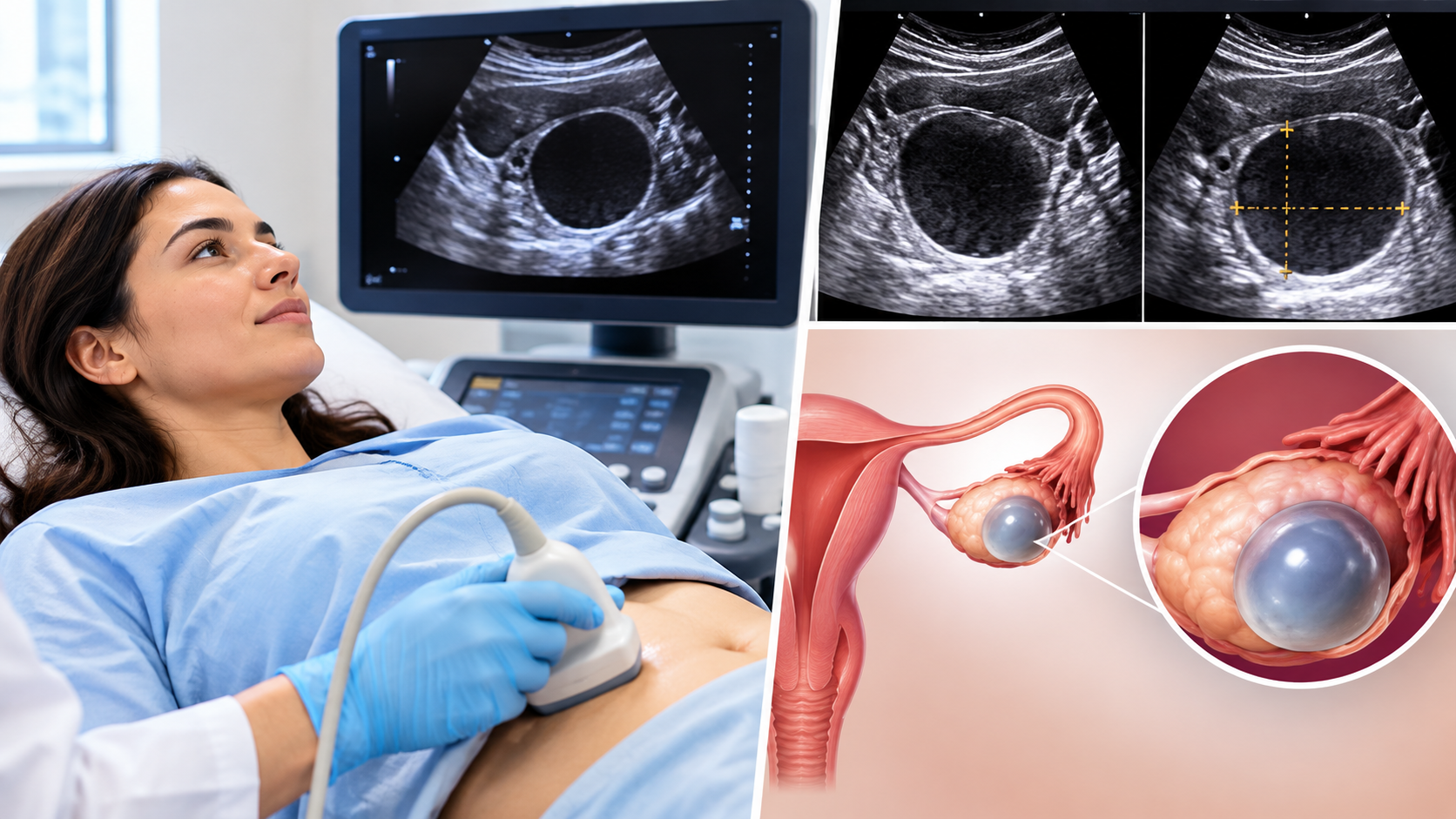
A simple cyst is the textbook benign finding: a smooth black circle.
Features radiologists list:
- Anechoic, meaning pure black with no internal echoes
- Thin, smooth wall
- Round or oval shape
- No septations (internal dividing walls)
- No solid parts or nodules
- Posterior acoustic enhancement, a bright band behind the cyst because sound passes through fluid easily
- No internal blood flow on Doppler
Most simple cysts are functional. A follicular cyst forms when a follicle grows but doesn’t release its egg. A corpus luteum cyst forms after ovulation when the empty follicle seals over and fills with fluid. Both are normal cycle events and most resolve within 8 to 12 weeks.
A simple cyst under 3 centimeters in a woman still having periods is a normal follicle. Many radiologists don’t even call it a cyst.
Complex Ovarian Cyst on Ultrasound
Complex means anything that isn’t simple. The label sounds alarming; it isn’t a diagnosis, just a description.
A complex cyst shows one or more of:
- Internal echoes (gray material inside the black fluid)
- Septations dividing the cyst into compartments
- A thick or irregular wall
- Solid areas or nodules on the wall
- Blood flow inside solid parts on color Doppler
Most complex cysts are still benign. Hemorrhagic cysts, endometriomas, and dermoid cysts are all technically complex, and all three have patterns experienced readers recognize on sight. The radiologist’s job is matching the pattern; your doctor’s job is deciding whether it needs follow-up.
Features that raise cancer concern are specific: solid tissue with blood flow, papillary projections (finger-like growths) larger than 3 millimeters, thick irregular septations, ascites, and a cyst larger than 10 centimeters. One of these is a reason for more testing, not a reason to panic.
Hemorrhagic Cyst on Ultrasound
A hemorrhagic cyst is a functional cyst that bled into itself, usually a corpus luteum. It often causes sudden one-sided pelvic pain around mid-cycle. We see this often enough that staff call it the “ER cyst”, patients frequently land in urgent care first, convinced something ruptured.
The pattern changes as the blood clots and breaks down:
- Early: a fine, lacy, net-like pattern of thin strands, called a reticular or fishnet pattern
- Later: a retracting clot — a gray blob with a concave edge sitting inside the fluid
- No blood flow inside the clot on Doppler
- A rim of strong blood flow around the wall, called the ring of fire, when it started as a corpus luteum
A hemorrhagic cyst under 5 centimeters in a premenopausal woman needs no follow-up. Larger ones get a repeat scan in 6 to 12 weeks to confirm resolution. In a postmenopausal woman, it’s unusual and gets a closer look.
Endometrioma on Ultrasound
An endometrioma forms from endometriosis tissue growing on the ovary and fills with old blood, giving it a distinct look:
- Homogeneous low-level echoes throughout, described as ground-glass
- Thick wall
- Usually no solid parts
- Sometimes small bright spots on the wall
- Little or no internal blood flow
Endometriomas don’t resolve on their own. They get a repeat scan in 6 to 12 weeks to confirm the diagnosis, then yearly monitoring if not removed.
They also tell you something about your body. A patient in her late 20s came to us after years of being told her pain was “just bad periods.” Her endometrioma on ultrasound was the first objective evidence anyone had shown her. Endometriosis is an estrogen-driven, inflammatory condition, and that ground-glass cyst is often the first hard proof of it.
What Does a Dermoid Ovarian Cyst Look Like on Ultrasound?
A dermoid cyst, also called a mature cystic teratoma, forms from germ cells and contains tissue like hair, fat, skin, and sometimes teeth. Benign in over 99 percent of cases. It doesn’t resolve on its own.
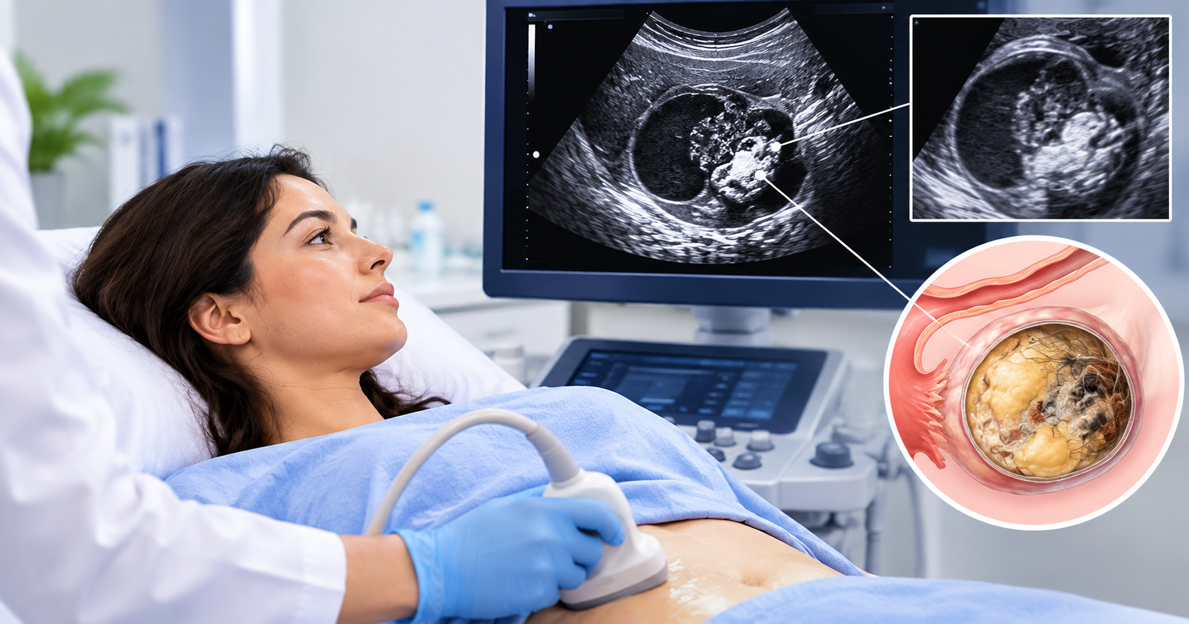
A dermoid cyst ovary on ultrasound is often the most memorable image a sonographer shows a patient: dense, textured, almost bone-white in places. Dermoids are the most variable cyst on ultrasound because their contents vary. Three signs point to the diagnosis.
Characteristic Ultrasound Signs of a Dermoid Cyst
- Rokitansky nodule. A bright, solid, shadowing lump on one wall — the plug where hair, teeth, and fat concentrate. Also called a dermoid plug.
- Tip of the Iceberg Sign. Fat and hair reflect sound so strongly that the waves can’t pass through. The front surface appears as a bright white band; everything behind is hidden in shadow. You see the tip, not the whole mass. The most recognized sign of a dermoid ovarian cyst on ultrasound.
Dermoid Mesh and Fat Fluid Level
- Dermoid mesh. Strands of hair floating in fluid create bright dots and dashes across the cyst — the dot-dash pattern.
- Fat fluid level. Fat floats on water. When a dermoid contains both, a flat horizontal line divides the bright fat layer above from darker fluid below. The line shifts if you change position.
Most dermoids are removed surgically; they grow slowly, carry a small torsion risk, and don’t go away. Small, symptom-free ones are sometimes watched with yearly ultrasound. One case that stuck with our team: a teenager whose dermoid was found incidentally during a scan for appendicitis no symptoms, just a Rokitansky nodule sitting quietly on a routine image.
How to Read Your Ovarian Cyst Ultrasound Results?
Your report follows a pattern. Here’s how to decode it.
- Location — Right or left adnexa (the area beside the uterus, including the ovary and tube).
- Size — Usually three measurements in centimeters, or a single largest diameter. Under 5 cm is small. 5 to 10 is moderate. Over 10 gets attention regardless of appearance.
- Contents — Anechoic means fluid, benign. Hypoechoic or low-level echoes mean blood or debris. Hyperechoic means fat, hair, or calcium.
- Wall and septations — Thin and smooth is reassuring. Thick, irregular, or nodular prompts follow-up.
- Solid components — Any mention of solid tissue, mural nodule, or papillary projection is worth asking your doctor about.
- Doppler — No flow or peripheral flow only is reassuring. Flow inside solid parts prompts more testing.
- Free fluid — A small amount is normal, especially after ovulation. A large amount, called ascites, is not.
- O-RADS score — Many reports now include an Ovarian Adnexal Reporting and Data System score from 1 to 5.
- O-RADS 1: normal ovary
- O-RADS 2: almost certainly benign (under 1 percent cancer risk)
- O-RADS 3: low risk (1 to 10 percent)
- O-RADS 4: intermediate risk (10 to 50 percent)
- O-RADS 5: high risk (over 50 percent)
Simple cysts, hemorrhagic cysts, endometriomas, and typical dermoids under 10 cm all score O-RADS 2.
- Impression — The radiologist’s conclusion and follow-up recommendation. Read this first.
When Is Further Testing Needed After an Ovarian Cyst Ultrasound?
Most cysts need one of three things: nothing, a repeat scan, or a referral.
Nothing further:
- Simple cyst up to 5 cm before menopause
- Simple cyst up to 1 cm after menopause
- Hemorrhagic cyst up to 5 cm before menopause
Repeat ultrasound:
- Simple cyst 5–7 cm before menopause, in 8 to 12 weeks
- Simple cyst over 1 cm after menopause, in 6 to 12 months
- Hemorrhagic cyst over 5 cm, in 6 to 12 weeks
- Any endometrioma, in 6 to 12 weeks, then yearly
- Any dermoid not removed, yearly
- Any cyst rated O-RADS 3
MRI:
- Simple cyst over 7 cm
- A cyst the ultrasound can’t characterize
- O-RADS 4 findings where surgery is being considered
Blood tests:
- CA-125 in postmenopausal women with a complex cyst, or in premenopausal women with suspicious features. CA-125 rises with endometriosis, fibroids, pelvic infection, and menstruation, so a high result in a premenopausal woman isn’t a cancer diagnosis on its own.
- Pregnancy test, since corpus luteum cysts are normal in early pregnancy
Referral to a gynecologic oncologist:
- O-RADS 4 or 5
- Solid components with blood flow
- Ascites
- Elevated CA-125 after menopause
Menopausal status changes the math. Before menopause, ovulation produces cysts every month, and most are functional. After menopause, the ovaries are quiet, so a new cyst has fewer innocent explanations. The same 4-centimeter simple cyst gets a shrug at 32 and a repeat scan at 62.
What Your Ultrasound Says About the Rest of Your Health?
An ovarian cyst on ultrasound is a snapshot of one organ and a data point about your hormones.
Repeated functional cysts often mean ovulation is irregular: a follicle grows, stalls, and never releases. This shows up with stress, thyroid dysfunction, insulin resistance, and perimenopausal hormone shifts. We’ve seen this in patients juggling high-stress jobs and cycles that never quite complete ovulation; the cyst on the scan was the visible tip of a larger hormonal picture.
A scan showing 20 or more small follicles on one ovary, or ovarian volume over 10 mL, meets the ultrasound criterion for polycystic ovaries. On its own, it doesn’t diagnose PCOS. Combined with irregular cycles or elevated androgens, it does. Learn how we assess and support PCOS:
An endometrioma points to estrogen excess and inflammation, both of which respond to root cause treatment alongside standard gynecologic care.
If your ultrasound came back benign and your doctor said watch and wait, use that time to look at why the cyst formed. A comprehensive hormone and metabolic assessment answers this question.
Frequently Asked Questions
Can ultrasound tell if an ovarian cyst is cancerous?
Ultrasound doesn’t diagnose cancer; it estimates risk. A simple cyst carries under 1 percent cancer risk. A cyst with solid parts, internal blood flow, thick septations, and pelvic fluid carries a much higher risk. The O-RADS score puts a number on it. Confirmation requires surgery and pathology.
What does a simple ovarian cyst look like on ultrasound?
A round black circle with a thin, smooth wall, no internal echoes, no septations, no solid parts, and no blood flow inside, with a bright band behind it. The most common finding and the most reassuring one.
How accurate is ultrasound for diagnosing ovarian cysts?
Transvaginal ultrasound correctly classifies about 90 percent of adnexal masses as benign or suspicious when read by an experienced operator. Accuracy is highest for simple cysts, endometriomas, and dermoids since their patterns are distinctive. It’s lower for complex cysts without a clear pattern, where MRI adds value.
What does a dermoid ovarian cyst look like on ultrasound?
A bright shadowing nodule on the wall, a bright front surface with shadow behind it (tip of the iceberg), floating dots and dashes from hair (dermoid mesh), and sometimes a flat line separating fat from fluid. Any one sign points to the diagnosis; two or more confirm it.
Do all ovarian cysts show up on ultrasound?
Nearly all cysts over 1 cm show up on transvaginal ultrasound. Very small cysts, cysts hidden behind bowel gas on a transabdominal scan, and cysts in women with a larger body habitus are sometimes missed. If symptoms persist and the scan is normal, a repeat transvaginal scan or MRI is next.
Take the Next Step for Your Health!
Your ultrasound report is a starting point. If it showed a simple or functional cyst, you likely need nothing more than a repeat scan and some attention to why your ovaries produced it. If it showed something complex, your gynecologist will guide the imaging and referral.
Either way, an ovarian cyst is a message about your hormones. Conscious Medicine works alongside your gynecologist to find out what your body is telling you and what to do about it. Bring your ultrasound report to your consultation, and we’ll go through it together.